Tracking down tumors with quantum optics
€6.7M project works on new tool for cancer diagnostics – Rapp Optoelectronic coordinates partners from industry and academia.
Quantum imaging enables insights into previously invisible areas. Can tumor diagnostics also benefit from this? This question is now being investigated by nine project partners, among them TU Darmstadt, which has particular expertise in quantum optics. The “Quancer” research project has a budget of 6.7 million euros and is being funded with 5.6 million euros by the German Federal Ministry of Education and Research (BMBF) under the framework program “Quantum Technologies – From the Basics to the Market”.
Various imaging techniques are used in the diagnosis of cancer. The aim is to detect tumor tissue and make it visible. When doctors discover suspicious tissue, they take a sample and examine it further: the gold standard of diagnostics are contrast methods that stain certain molecules and light microscopy to show their distribution.
Increasingly, digital microscopes are being used, which enable automated procedures and thus faster processes. Infrared-based imaging techniques, such as infrared microscopy, tie in with digital pathology and provide further information. To do this, infrared light is used to excite molecules. Based on the vibrations of the molecules, it is now possible to infer their nature. Tissue is thus made visible without the need for additional contrast agents. However, this method faces limitations in detection because infrared detectors are limited in efficiency and signal-to-noise ratio.
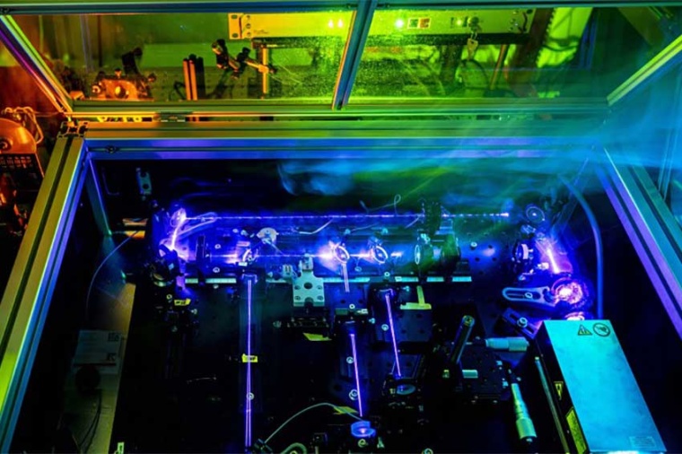
Quantum imaging can be used to circumvent this problem. This is done by using two light beams correlated with each other in a specific arrangement. In simplified terms, one light beam thereby sends photons, or particles of light, to the tissue sample. The other light beam sends photons to a camera. Due to the quantum correlation of both photons, an image of the tissue sample is generated, although the light reaching the camera has never “seen” it – this is “spooky” quantum imaging.
Quantum imaging is now being combined with a professional microscopy system for the first time and will be tested in a clinical setting as part of the project. “At TU Darmstadt, we take care of most of the experimental work of a fundamental nature,” explains Markus Gräfe, professor at the Institute of Applied Physics. “So we build the first laboratory experiments that show that everything works as desired and with which we investigate and optimize certain quantum imaging modes. Our setups are then converted into compact forms by the Fraunhofer IOF and combined with the microscope developed by our industrial partner Rapp Optoelectronic. The system will then be used at the Jena University Hospital. In perspective, this will introduce a new tool for cancer diagnostics.”
Further project partners are Leibniz IPHT, Toptica Photonics, Institute of Applied Physics at Friedrich Schiller University Jena, Institute for Laser Physics at Hamburg University, and n-Hands.