New fluorescence imaging system for fast light intensity measurements
04.01.2024 - Two new complementary approaches for versatile but precise light intensity measurements.
An international collaboration of researchers developed two new complementary approaches for versatile but precise light intensity measurements in fluorescence imaging systems. Led by Ludovic Jullien from École Normale Supérieure, Sorbonne University, and CNRS, the research team used organic dyes and proteins to develop two new fluorescence-based methods for the holistic but precise measurement of light intensity, a growing need for many scientists. The collaboration involved researchers from several CNRS groups and partners of the EIC-funded DREAM project, including the Forschungszentrum Jülich, Sony Computer Science Laboratories, and Palacký University. These protocols also proved effective for calibrating commercial instruments and light sources.
-
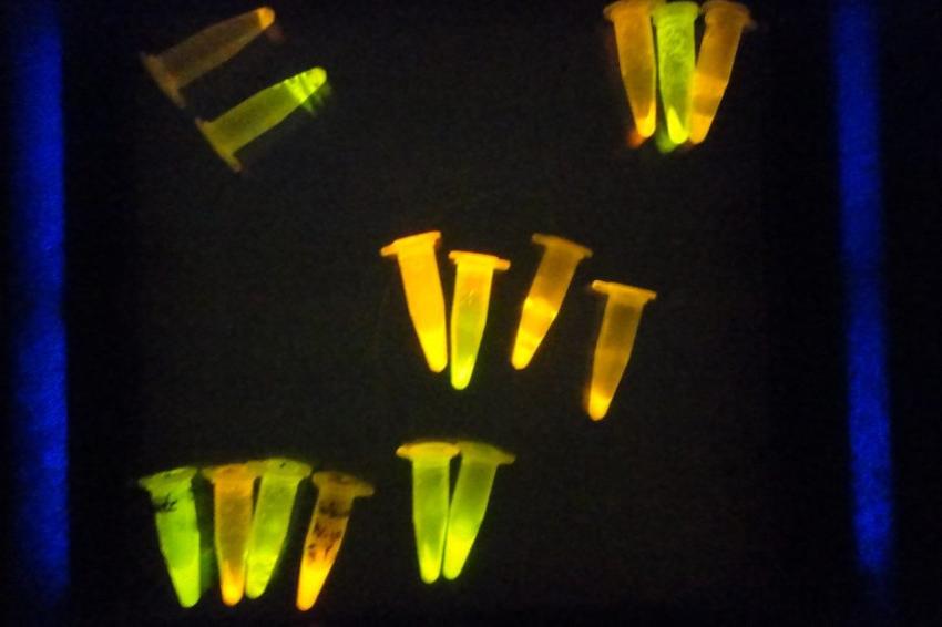 Green fluorescent proteins in eppendorf tubes glow green because they have absorbed exitation light. (Source: J. C. F. Mata)
Accurate measurements of light intensity provide vital data for scientists and everyday applications. For example, precise values help optimize microscopy signals, trigger physiological processes in the brain, and drive light-absorbing reactions while enabling different research teams to share and reproduce experimental results. “Nowadays, a vast community of biologists, chemists, engineers, and physicists are concerned with delivering precise numbers of photons,” the team explains. On a larger scale, precision is also essential for critical tasks such as water purification and phototherapeutics.
However, most approaches lack versatility and usability. For example, most methods cannot quantify light intensity and spatial distribution simultaneously or can only do so across a limited range of light wavelengths and intensities. To provide a versatile alternative to the limitations of current approaches, the research team developed two complementary protocols using novel actinometers – physical or chemical systems that can quantify photons. As they are liquid solutions, actinometers can be used in samples of different shapes and sizes. Still, most are not very accurate or compatible with imaging systems and are usually limited to specific wavelengths and light intensities.
To overcome these limitations, the team used fluorescence-based actinometers, which proved to be faster, more sensitive, and able to provide data that is more accessible to imaging systems. The first protocol uses five molecular actinometers – covering the entire spectrum of ultraviolet and visible light – that emit fluorescent signals when they absorb the excitation light that the researchers want to measure. Under certain conditions, this protocol can also map the spatial distribution of light intensity. The team tested several types of actinometers, from synthetic chemicals for chemists to proteins and photosynthetic organisms for biologists. “We wanted to make fluorescent actinometers accessible to different communities of end users,” the researchers mentioned.
The second protocol complements the fluorescent actinometers of the first because, due to their limited ranges of light absorption, several actinometers are needed to cover the whole range of wavelengths. This protocol uses a stable fluorophore–a chemical that can re-emit light upon light excitation–to back-calculate light intensity from one wavelength to another. “Together, the two new protocols can be used in weak-light situations, shorter periods, and a wider range of wavelengths than conventional methods,” affirmed the group.
The research team successfully applied the protocols to characterize the spatial distribution of light in different fluorescence imaging systems, demonstrating their versatility and accuracy. The protocols have also been used to calibrate illumination in commercially available instruments and light sources.These protocols could be applied in wide-field and confocal microscopy, enabling precise measurement of light intensity in biological samples. “We envision that [our protocols] will improve our understanding of how light affects the health and viability of biological specimens.” Even complex environments can be studied, such as within gels or deep within tissues, an essential feature for DREAM in its mission to record the dynamics of photosynthesis regulation in microalgae or plants.
Introducing these fluorescence-based actinometry methods represents a significant leap forward in accurately measuring light intensity. With online access to actinometer properties and user-friendly data processing applications, researchers and engineers should now be equipped to accurately measure light intensity and share reliable and reproducible data across disciplines. (Source: Insociety)