Intelligent microscopes detect biological events
New control software optimizes how fluorescence microscopes collect data on living samples
Biophysicists from the Swiss Federal Institute of Technology in Lausanne have found a way to automate microscope control for imaging biological events in detail while limiting stress on the sample, all with the help of artificial neural networks. Their technique works for bacterial cell division, and for mitochondrial division. “An intelligent microscope is kind of like a self-driving car. It needs to process certain types of information, subtle patterns that it then responds to by changing its behavior,” explains principal investigator Suliana Manley of the Laboratory of Experimental Biophysics. “By using a neural network, we can detect much more subtle events and use them to drive changes in acquisition speed.”
-
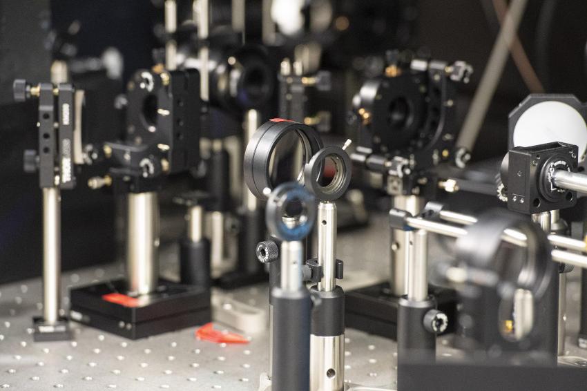 Swiss biophysicists have developed control software that optimizes how fluorescence microscopes collect data on living samples. (Source: EPFL)
Manley and her colleagues first solved how to detect mitochondrial division, more difficult than for bacteria such as C. crescentus. Mitochondrial division is unpredictable, since it occurs infrequently, and can happen almost anywhere within the mitochondrial network at any moment. But the scientists solved the problem by training the neural network to look out for mitochondrial constrictions, a change in shape of mitochondria that leads to division, combined with observations of a protein known to be enriched at sites of division.
When both constrictions and protein levels are high, the microscope switches into high-speed imaging to capture many images of division events in detail. When constriction and protein levels are low, the microscope then switches to low-speed imaging to avoid exposing the sample to excessive light. With this intelligent fluorescent microscope, the scientists showed that they could observe the sample for longer compared to standard fast imaging. While the sample was more stressed compared to standard slow imaging, they were able to obtain more meaningful data.
“The potential of intelligent microscopy includes measuring what standard acquisitions would miss,” Manley explains. “We capture more events, measure smaller constrictions, and can follow each division in greater detail.” The scientists are making the control framework available as an open source plug-in for the open microscope software Micro-Manager, with the aim of allowing other scientists to integrate artificial intelligence into their own microscopes. (Source: EPFL)