Tiny endo-microscope for deep-brain observations
New device could help to understand neuronal communication better.
In order to investigate the activity of neuronal structures as well as the interaction of nerve cells, minimally invasive technologies providing images from delicate deep-brain tissues are required. A new hair-thin endo-microscope, developed by an international team with the participation of Leibniz Institute of Photonic Technology (Leibniz IPHT) in Jena, Germany, promises extremely gentle in-depth observations. It offers the potential to investigate areas of the brain in great detail and to study the onset and progression of severe neuronal diseases. The instrument is expected to help neuroscientists in defining new strategies how to combat these debilitating conditions.
Neuronal diseases such as autism, epilepsy, Alzheimer’s, or Parkinson’s disease are still poorly understood. Therefore, preventing, treating, or alleviating these illnesses remains a major challenge. In order to better understand the causes and the origins of these diseases, and to develop and monitor tailored therapies, it is important to decipher and study how the affected nerve cells, which are frequently located in very deep structures of the brain, behave within the natural complexity of the whole organism. Neuroscientists investigate these conditions in small animal models and exploit minimally invasive endoscopy techniques to study deep-brain structures.
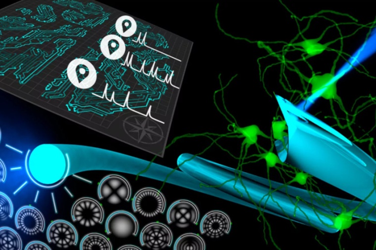
With a diameter of only 110 micrometers, the developed endoscope is enabling the acquisition of images at an unprecedented tissue depth and at the subcellular level. This not only allows research into deep-residing brain structures, which were previously difficult to access, but also to precisely study the neuronal connectivity and signaling activity of individual neurons in the brain. “At the heart of the endoscopic system is an ultra-thin optical glass fiber that serves as a probe. Using digital holography, we can use it to image and visualize individual cells and blood vessels with high-resolution, distortion-free and with high-contrast. The hair-thin endoscopy design enables extremely atraumatic in-vivo examination without damaging surrounding tissues,” explains Tomáš Čižmár, head of the Fiber Research and Technology Research Department at Leibniz IPHT.
Conventional endoscopic solutions used in in-vivo neuroscience research usually use specialized rod lenses (GRIN), which convey images from one of its ends to the other. Due to their footprint, they may pose a great risk of tissue damage and severely affect the validity of neuroscience studies. The newly developed holographic endoscope overcomes this disadvantage by using a single multimode optical fiber as the imaging probe, making it the least invasive method for visualizing sensitive brain areas.
The researchers have also exploited a new type of fiber probe, a side-view probe, which allows observation of the tissue perpendicular to the fiber axis. In this way, the tissue being imaged is much less stressed and damaged by the insertion of the endoscope into the tissue than by using conventional bare-terminated probes. As the probe offers the potential to penetrate deeper into the tissue, the side-view probe also offers the possibility to stitch together individual frames taken along the way into a panorama-like image, allowing a continuous view of the entire depth of the brain.
In addition to the direct observation of structural connectivity between neurons, particularly the dendritic spines, intracellular processes can also be observed with the developed endoscopic solutions. Dynamics of subcellular structures, changes in intra-cellular calcium concentration, and the speed of the blood flow in individual vessels could be studied. All these properties could provide neuroscientists with indications to pathological changes in the brain.
„In neuronal diseases, the cognitive and motor performance of the affected organism may be irreversibly limited by changes or loss of nerve cells. We are developing technologies to monitor signs of a disease, for example, through altered neuronal communication, at an early stage. With these new and powerful light-based instruments, we can help to provide unprecedented insights into the control center of vital functions with high image quality and thus expand the understanding of neuronal diseases,“ says Čižmár. (Source: Leibniz-IPHT)
Link: Fiber Research and Technology, Leibniz Institute of Photonic Technology, Jena, Germany