A flexible microdisplay visualizes brain activity in real time
10.05.2024 - The device represents a leap ahead to monitor brain activity to guide neurosurgeons.
A thin film that combines an electrode grid and LEDs can both track and produce a visual representation of the brain’s activity in real-time during surgery – an improvement over the current state of the art. The device is designed to provide neurosurgeons visual information about a patient’s brain to monitor brain states during surgical interventions to remove brain lesions including tumors and epileptic tissue.
-
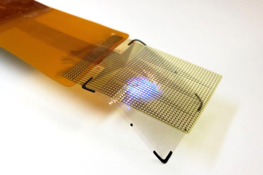 The device's LEDs can light up in several colors. This allows surgeons to see which areas they need to operate on and to track brain states during surgery. (Source: UCSD)
Each LED in the device mirrors the activity of a few thousand neurons. In a series of proof-of-concept experiments in rodents and large non-primate mammals, researchers showed that the device can effectively track and display neural activity in the brain corresponding to different areas of the body. In this case, the LEDs developed by the team light up red in the areas that need to be removed by the surgeon. Surrounding areas that control critical functions and should be avoided show up in green.
The study also showed that the device can visualize the onset and map the propagation of an epileptic seizure on the surface of the brain. This would allow physicians to isolate the nodes of the brain that are involved in epilepsy. It also would allow physicians to deliver necessary treatment by removing tissue or by using electrical pulses to stimulate the brain. “Neurosurgeons could see and stop a seizure before it spreads, view what brain areas are involved in different cognitive processes, and visualize the functional extent of tumor spread. This work will provide a powerful tool for the difficult task of removing a tumor from the most sensitive brain areas,” said Daniel Cleary, a neurosurgeon at Oregon Health and Science University. Cleary was a medical resident and a postdoctoral researcher at the University of California San Diego.
The device was conceived and developed by a team of engineers and physicians from University of California San Diego and Massachusetts General Hospital (MGH) and was led by Shadi Dayeh. During brain surgery, physicians need to map brain function to define which areas of the organ control critical functions and can’t be removed. Currently, neurosurgeons work with a team of electrophysiologists during the procedure. But that team and their monitoring equipment are located in a different part of the operating room. Brain areas that need to be protected and those that need to be operated on are either marked by electrophysiologists on a paper that is brought to the surgeon or communicated verbally to the surgeon, who then places sterile papers on the brain surface to mark these regions. “Both are inefficient ways of communicating critical information during a procedure, and could impact its outcomes,” said Angelique Paulk of MGH, a co-inventor of the technology.
In addition, the electrodes currently used to monitor brain activity during surgery do not produce detailed fine grained data. So surgeons need to keep a buffer zone, known as resection margin, of 5 to 7 millimeters around the area they are removing inside the brain. This means that they might leave some harmful tissue in. The new device provides a level of detail that would shrink this buffer zone to less than a millimeter. “We invented the brain microdisplay to display with precision critical cortical boundaries and to guide neurosurgery in a cost-effective device that simplifies and reduces the time of brain mapping procedures,” said Shadi Dayeh.
Researchers installed the LEDs on top of another innovation from the Dayeh lab, the platinum nanorod electrode grid (PtNRGrid). Using the PtNRGrids since 2019, Dayeh’s team pioneered human brain and spinal cord mapping with thousands of channels to monitor brain neural activity. The PtNRGrid also includes perforations, which enable physicians to insert probes to stimulate the brain with electrical signals, both for mapping and for therapy.
Dayeh and his team used their expertise in working with gallium nitride to develop a manufacturing technique for high-efficiency LEDs that do not heat up when they light up and do not damage brain tissues. The material itself is grown on a flat and rigid substrate called Qromis substrate technology. Dayeh’s team was able to embed thousands of LEDs in flexible films and release them from the substrate in the form of a flexible display panel. Researchers then used inkjet printing to deposit quantum dot inks on the surface of the LEDs to convert their blue light to multiple other colors. “This enables richer and more nuanced visual representation of neural activity patterns,” said Dayeh.
“These gallium nitride-based inorganic micro-LEDs, substantially brighter and power-efficient than organic LEDs, can maintain clear visibility under surgical lights that may exceed the brightness of direct sunlight. The iEEG microdisplay, just a few tens of microns thick, captures brain activity at 20,000 samples per second across thousands of channels and visualizes it at a video rate of 40 Hertz. This enables precise and real-time displays of cortical dynamics during critical surgical interventions,” said Youngbin Tchoe, formerly a postdoc in the Dayeh group.
The microdisplays measure 5 by 5 square millimeters and 32 by 32 square millimeters and include either 1024 or 2048 of these LEDs, laminated on the back of the PtNRGrid. In addition to the LEDs, the device includes acquisition and control electronics as well as software drivers to analyze and project cortical activity directly from the surface of the brain. “The brain iEEG-microdisplay can impressively both record the activity of the brain to a very fine degree and display this activity for a neurosurgeon to use in the course of surgery. We hope that this device will ultimately lead to better clinical outcomes for patients with its ability to both reveal and communicate the detailed activity of the underlying brain during surgery,” said neurosurgeon Jimmy Yang at Ohio State University.
Dayeh’s team is working to build a microdisplay that will include 100,000 LEDs, with a resolution equivalent to that of a smartphone screen. Each LED in those displays would reflect the activity of a few hundred neurons. These brain microdisplays will cost a fraction of a high-end smartphone. This brain microdisplay will also include a foldable portion. This would allow surgeons to operate within the foldable portion and monitor the impact of the procedure as the other, unfolded portion of the microdisplay shows the status of the brain in real time.
Researchers are also working on one limitation of the study. The close proximity of the LED sensors and the PtNRGrids led to a slight interference and noise in the data. The team plans to build customized hardware to change the frequency of the pulses that turn on the LEDs to make it easier to screen out that signal, which is not relevant to the brain’s electrical activity. (Source: UCSD)