Novel imaging system for near-instant biopsy results
02.11.2022 - New technology could enable point-of-care skin cancer diagnosis.
Medicine has advanced dramatically during the last century. But when it comes to getting biopsy results, very little has changed. Consider, for example, what happens when a patient comes in to have a skin lesion biopsied for nonmelanoma skin cancer. “The surgeon will take a little piece of the skin out,” says Michael Giacomelli, an assistant professor of biomedical engineering and of optics at the University of Rochester. Next, “someone in pathology will look at it under a microscope. And then, depending on what they find, the patient is notified that everything’s fine, don’t worry about it, or we need you to come back for a second appointment so we can treat you.”
-
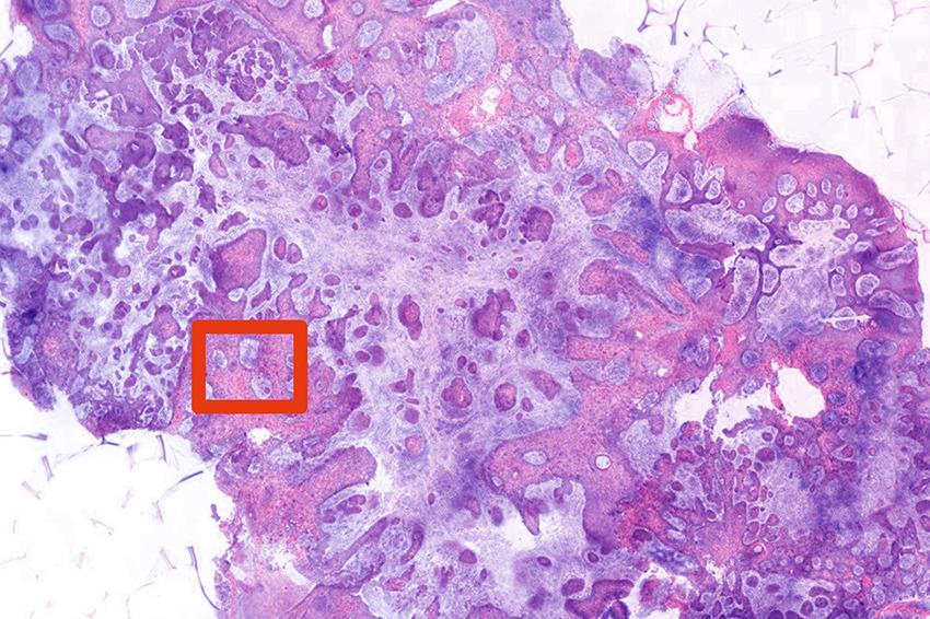 Tissue biopsied with a novel imaging system based on 2-photon fluorescence microscopy (TPFM) is showing promising results. (Source: Giacomelli lab, U. Rochester)
Giacomelli is developing a novel imaging system, contained on a portable cart, to shorten this process to two minutes. This would enable a surgeon to immediately determine whether the lesion is cancerous and, if so, to “treat the patient during the same visit instead of stretching it out over the next month and multiple visits.” The system –using two-photon fluorescence microscopy (TPFM) – demonstrated remarkable accuracy in a pilot study. When tested on 15 biopsies of known nonmelanoma skin cancer, the technology was able to detect basal cell carcinoma with perfect accuracy – 100 percent sensitivity and specificity – and squamous cell carcinoma with high accuracy – 89 percent sensitivity and 100 percent specificity.
Nonmelanoma skin cancer is the most common type of human cancer, with more cases annually than all other types of cancer combined in the United States. Eighty percent of nonmelanoma skin cancers are basal cell carcinoma. Typically biopsied tissue is excised, fixed, paraffinized(preserved), stained, and mounted on slides before being evaluated by a dermatopathologist. “This is how it has been done since the late 19th century,” Giacomelli says. “Billions and billions of biopsies have been done this way. Everybody is on the same page; it works great. The problem is that it is very slow.”
Even when tissue is quickly frozen for quicker analysis, surgeons can wait an hour or more for the biopsy results to be sure they have completely removed a malignant tumor from a patient still on the operating table. This ties up the surgeon’s time and the operating room, while interrupting the workflow in busy pathology departments. Giacomelli sees potential applications for his system in quickly providing biopsy results for all kinds of diseases. “There are all sorts of scenarios,” he says.
For example, “in prostate cases, surgeons often have a very poor idea what they’re getting into based on just an X-ray or MRI image beforehand,” Giacomelli says. “You see random cases where they take out the prostate, and sometimes there’s an obvious tumor that has spread beyond the prostate, or other times it is very localized. This technology offers a possibility to adjust the patient’s therapy on the fly.”
The advantage of using TPFM is that it not only generates high-resolution images but it also uses near-infrared light that penetrates deeper through tissue, making it “advantageous for rapid imaging of fresh, irregularly shaped biopsies with minimal preparation,” the researchers note. Giacomelli is now working closely with Sherrif Ibrahim, an associate professor of dermatology at the University’s Medical Center, on a larger, 200-patient follow up study, using biopsy samples taken at random at Ibrahim’s Rochester Dermatological Surgery in nearby Victor, New York.
“The goal is to analyze how well the technique works in a real-world scenario, where you have a lot people coming in and all kinds of things can show up,” Giacomelli says. “We really want to make sure that there isn’t some bizarre thing that normally has nothing to do with cancer that we somehow confuse with cancer – or something that does have something to do with cancer but we can’t image. You never know what you’re going to find once you start taking random biopsies of people.
Giacomelli has also embarked on a parallel study to see if a novel system he has developed incorporating TPFM and video can be used to guide surgeries. The system overlays TPFM images of the site where tissue is being removed on a webcam image, co-registering what it looks like to the eye with what it would look like processed on a slide. “You can show what the excised tissue looks like, and how much of the tumor is located on the excision,” Giacomelli says. “They can then map that back into the wound that they are creating in order to decide which side to cut.” (Source: U. Rochester)
Link: Dept. of Biomedical Engineering, University of Rochester, Rochester, New York